| Age group | Applied modality | Imaging signs | |
| With primary malformation(n=22) | With secondary malformation(n = 14) | ||
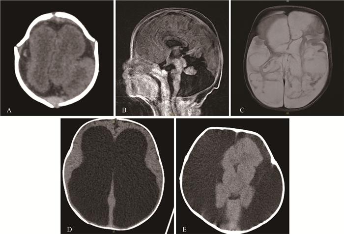
| ≤ 1 year | MRI(n=3) CT(n=16) | Lissencephaly(n=1),Dandy-walker and corpus callosum hypoplasia(n=1),Partial absence of gyrus(n=2),Polygyria(n=1),Cystic deformation of cerebral parenchyma(n=2) | Communicative hydrocephalus(n=3), Encephalatrophy(n=7), Porencephaly(n=2) |
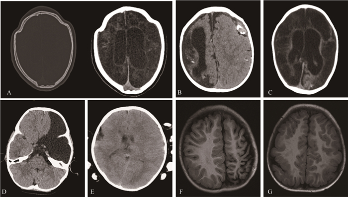
| 1-3 years | CT(n=7) | Craniosynostosis(n=2),Polygyria(n=2),Cystic deformation of cerebral parenchyma(n=3) | - |
| >3 years | MRI(n=4) CT(n=6) | Partial absence of gyrus(n=1),Polygyria or pachygyria(n=4),Dysplasia of the gyrus(n=2),Porencephaly(n=1) | Ventriculomegaly(n=1), Encephalatrophy(n=1) |
Fig 1 Imaging demonstrations of CNS malformations in the ≤ 1-year-old group A: CT revealed lissencephaly in a one-month-old infant; B: Dandy-walker malformation was found on sagittal MRI in a 1-month-old infant who died 3 months after birth; C: The cystic changes in bilateral cerebral parenchyma was found on MRI in a 2-month-old infant; D: The heavy hydrocephalus was found on CT in a 2-month-old infant who died after one-month treatment; E: The extent subdural effusion was found on CT for a 10-month-old infant who died one year after birth.
Fig 2 Imaging demonstrations of CNS malformations in the 1- to 3-year-old group and>3-year-old group A: The irregular contour of the cranium suggests craniosynostosis in a 24-month-old infant (left) and the relatively low density in both cerebral parenchyma was found(right); B: The dysplasia of gyrus in the right hemisphere and the corresponding porencephaly was found on CT image in a 24-month-old infant. The high-density hematoma was also found in the contralateral subdural space; C: The CT indicated the extent low density of both cerebral parenchyma in a 36-month-old infant. D: The absence of left frontal lobe in axial CT was found in a 48-month-old child. The subsequent follow-up demonstrated the barylalia with normal movement function; E: The giant gyrus in the right and the polygyria in left hemisphere on CT was found in a 60-month-old child. F: The dysplasia of left both frontal and parietal lobe was found on MRI in a 48-month-old infant. G: The polygyria was found in both the bilateral frontal and parietal lobe on axial MRI in a 48-month-old infant.