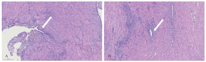
Fig 1 Transvaginal ultrasonography of uterine and bilateral appendages A: The shape of uterine cavity was roughly normal, no abnormalities were found in bilateral uterine corners, and internal endometrial echo was observed, with thickness of about 12 mm; B: An uneven echo block with a size of 4.5 cm×3.9 cm was visible below the right fundal angle, with clear boundaries, regular shape and ground-glass like liquid echo.
Fig 2 Results of laparoscopic examination A: The uterus was slightly full under laparoscopy, and no obvious abnormalities were observed. A mass with a diameter of 4 cm was visible onthe right anterior wall of the uterus, located below the anterior circular ligament of the right side, and no obvious abnormalities were observed inthe bilateral accessory area. The arrow indicates the position of the mass. B: In the process of removing the mass, the chocolate liquid was found toflow out. The arrow indicates the chocolate-like liquid. C: Uterine morphology was generally normal after operation.