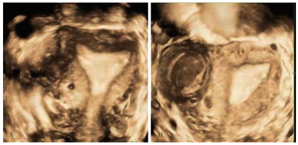
Fig 1 Transvaginal ultrasonography of uterine and bilateral appendages A: The shape of uterine cavity was roughly normal, no abnormalities were found in bilateral uterine corners, and internal endometrial echo was observed, with thickness of about 12 mm; B: An uneven echo block with a size of 4.5 cm×3.9 cm was visible below the right fundal angle, with clear boundaries, regular shape and ground-glass like liquid echo.
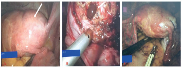
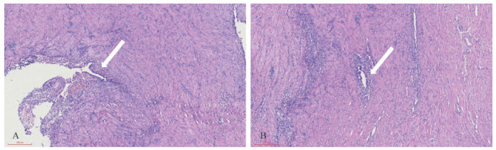
Other figure/table from this article