×
模态框(Modal)标题
在这里添加一些文本
Close
Close
Submit
Cancel
Confirm
×
模态框(Modal)标题
×
Welcome to visit Fudan University Journal of Medical Sciences,
Share:
Toggle navigation
Home
About
About Journal
Honor
Indexed In
Editorial Board
Editorial Board
Youth Editorial Board
Instruction
FAQ
Journal Online
Just Accepted
Current Issue
Archive
Most Read
Most Download
Most Cited
E-mail Alert
Download
Journal Policy
Academic Publishing Standards
Ethical Policies
Copyright Policy
Review Process
Correction and Withdrawal
Others
Contact Us
中文
Figure/Table detail
TAFRO syndrome: one case report and literature review
Man-man LI, Yun-hua HOU, Chen-chen WANG, Ming DING, Xiao-xiao WANG, Zheng WEI
Fudan University Journal of Medical Sciences
, 2025, 52(
02
): 305-310. DOI:
10.3969/j.issn.1672-8467.2025.02.020
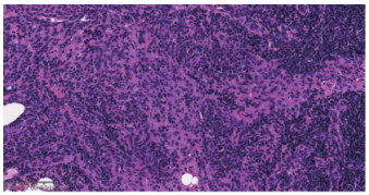
Fig 5
Lymph node biopsy shows patchy distribution of plasma cells under high magnification (HE×100)
Other figure/table from this article
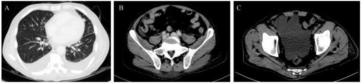
Fig 1
Chest and abdominal CT shows a small amount of bilateral pleural effusion, a small amount of pericardial effusion,
A: Bilateral pleural effusions (right-sided depth: 3.7 cm, left-sided: 2.6 cm) with minimal pericardial effusion (fluid layer thickness: 0.7 cm); B: Ascites (scattered fluid-density opacities in the peritoneal cavity); C: Pelvic effusion (patchy fluid-density opacities with maximal measurements: 11.6 cm×7.9 cm). a small amount of abdominal effusion and a big amount of pelvic effusion
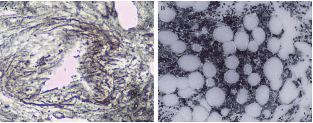
Fig 2
Temporal pathological changes of bone marrow fibrosis
A: On the first day of admission to the hematology ward, bone marrow biopsy showed proliferation of bone marrow fibrous tissue (reticular fiber staining×40: MF1-MF2); B: On the 29th day of admission to the hematology ward, bone marrow biopsy showed no proliferation of bone marrow fibrous tissue (reticular fiber staining×10: negative).
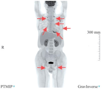
Fig 3
PET-CT shows enlarged lymph nodes with multiple increased FDG uptake throughout the body
FDG:
18
F-Fluorodeoxyglucose. Arrows indicated lymph nodes with increased FDG metabolism.
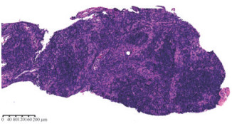
Fig 4
Lymph node biopsy specimen under low magnification
Lymph node biopsy shows nodular distribution of lymphocytes at low magnification, atrophy of the germinal center, proliferation of cells in the mantle and marginal regions, and proliferation of blood vessels in the stroma, with some glassy changes (HE×10).