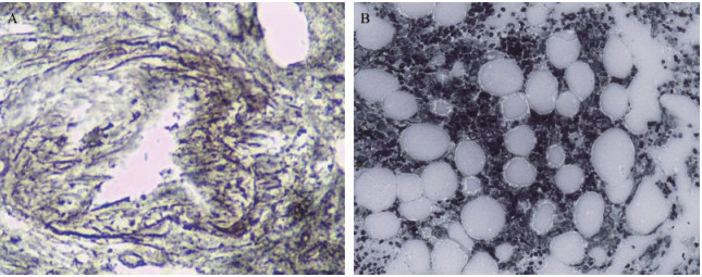
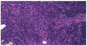
Fig 2 Temporal pathological changes of bone marrow fibrosis A: On the first day of admission to the hematology ward, bone marrow biopsy showed proliferation of bone marrow fibrous tissue (reticular fiber staining×40: MF1-MF2); B: On the 29th day of admission to the hematology ward, bone marrow biopsy showed no proliferation of bone marrow fibrous tissue (reticular fiber staining×10: negative).
Other figure/table from this article