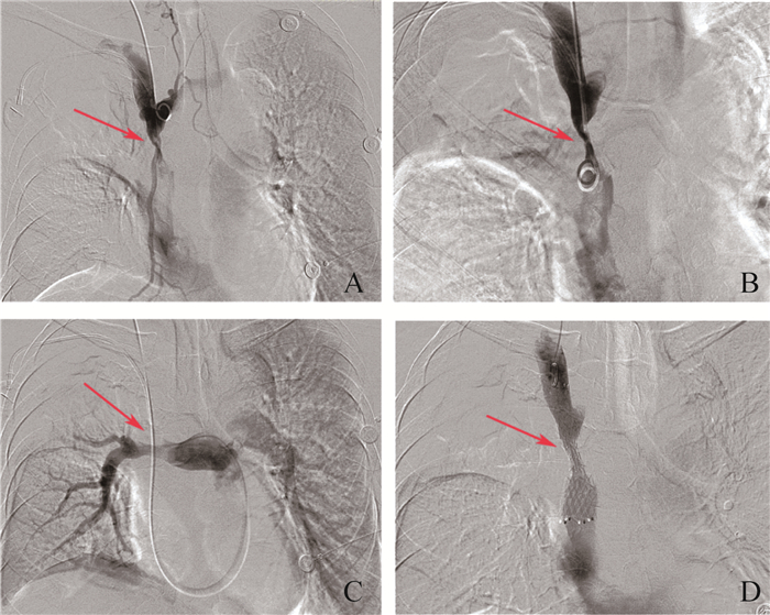
Fig 2 DSA images of superior vena cava before and after treatment A and B: Superior vena cava stenosis with collateral circulation; C: Stenosis of the right main pulmonary artery, the branches of right superior pulmonary artery were not visualized; D: Post-operative angiography showed that unobstructed blood flow in the superior vena cava. The red arrow in A, B and C showed the narrow segment, while the red arrow showed the placement of the stent in the superior vena cava.
Other figure/table from this article