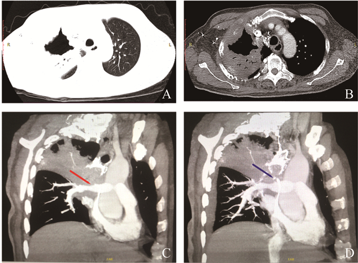
Fig 1 Chest CT and pulmonary artery CTA before treatment The lung window (A) and mediastinal window (B) of the axial HRCT showed a soft tissue shadow around the upper lobe of the right lung; C: Sagittal oblique CTA image of the PA showed stenosis at the origin of the right PA (the red arrow); D: A severe stenosis of the SVC (the blue arrow) and a large amount of collateral circulation around SVC. SVC: Superior vena cava; PA: Pulmonary artery.
Other figure/table from this article