×
模态框(Modal)标题
在这里添加一些文本
Close
Close
Submit
Cancel
Confirm
×
模态框(Modal)标题
×
Welcome to visit Fudan University Journal of Medical Sciences,
Share:
Toggle navigation
Home
About
About Journal
Honor
Indexed In
Editorial Board
Editorial Board
Youth Editorial Board
Instruction
FAQ
Journal Online
Just Accepted
Current Issue
Archive
Most Read
Most Download
Most Cited
E-mail Alert
Download
Journal Policy
Academic Publishing Standards
Ethical Policies
Copyright Policy
Review Process
Correction and Withdrawal
Others
Contact Us
中文
Figure/Table detail
Clinical efficacy observation of vital pulp therapy for mature permanent teeth with carious irreversible pulpitis
Zhi-ming QIN, Jia-yang LI, Hua-xing XU, Zhi-fei MA, Xiao-ling WEI
Fudan University Journal of Medical Sciences
, 2025, 52(
02
): 263-269. DOI:
10.3969/j.issn.1672-8467.2025.02.013
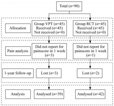
Fig 2
Consort flowchart of patients throughout the trial
VPT: Vital pulp therapy; RCT: Root canal treatment.
Other figure/table from this article
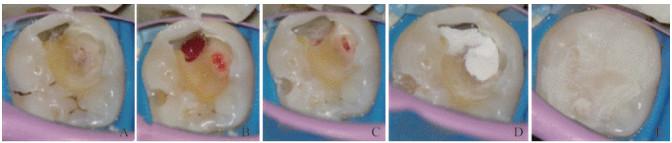
Fig 1
Examples of VPT procedures
A: Complete removal of carious tissues; B: Extirpation of the infected dental pulp; C: Gentle pressure for hemostasis using a sodium hypochlorite-soaked cotton ball; D: Coverage of the pulp stump with iROOT BP Plus; E: Adhesive restoration with composite resin.
Tab 1
Patient clinical data
[
n
(%) or $\bar x \pm s$]
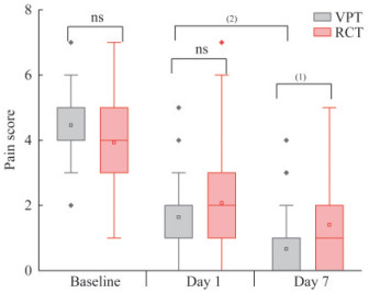
Fig 3
Pain intensity in VPT and RCT groups
(1)
P
< 0.01,
(2)
P
< 0.001, ns: No significance.
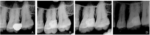
Fig 4
Preoperative and postoperative X-rays of case 1
A: Preoperative X-ray showed a high-density shadow of filling material on 26 occlusal and distal surface; B: Postoperative X-ray at 1 month; C: Postoperative X-ray at 6 month; D: Postoperative X-ray at 1 year.
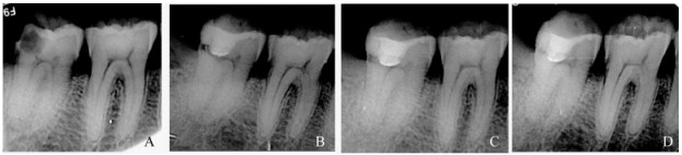
Fig 5
Preoperative and postoperative X-rays of case 2
A: Preoperative X-ray showed a low-density shadow of filling material on 47 occlusal and distal surface; B: Postoperative X-ray at 1 month; C: Postoperative X-ray at 6 month; D: Postoperative X-ray at 1 year.