×
模态框(Modal)标题
在这里添加一些文本
Close
Close
Submit
Cancel
Confirm
×
模态框(Modal)标题
×
Welcome to visit Fudan University Journal of Medical Sciences,
Share:
Toggle navigation
Home
About
About Journal
Honor
Indexed In
Editorial Board
Editorial Board
Youth Editorial Board
Instruction
FAQ
Journal Online
Just Accepted
Current Issue
Archive
Most Read
Most Download
Most Cited
E-mail Alert
Download
Journal Policy
Academic Publishing Standards
Ethical Policies
Copyright Policy
Review Process
Correction and Withdrawal
Others
Contact Us
中文
Figure/Table detail
Reference ranges of cardiac size and morphology for low-risk fetuses at 28-39 gestational weeks based on two-dimensional speckle tracking technique
Chen ZHU, Cheng-jie XU, Rui LIU, Man LI, Yu XIONG, Jin-lian XIANG, Yun-yun REN
Fudan University Journal of Medical Sciences
, 2024, 51(
01
): 41-49. DOI:
10.3969/j.issn.1672-8467.2024.01.006
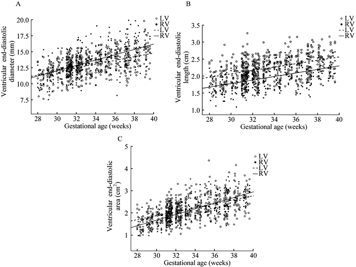
Fig 3
Scatterplots of ventricular size with gestational weeks in low-risk fetuses
Other figure/table from this article
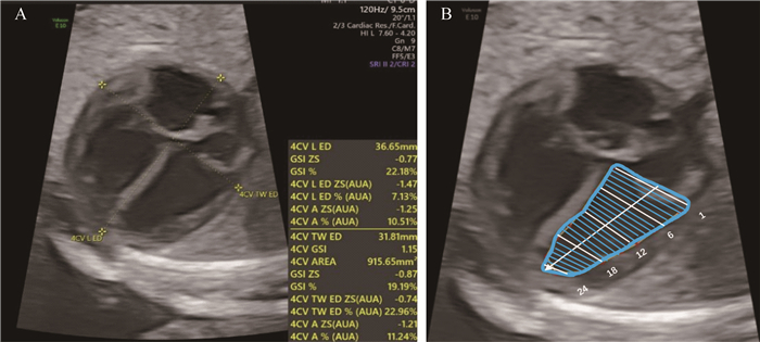
Fig 1
Measurement of fetal cardiac size and morphology
A: Measurement of four-chamber view end-diastolic size and sphericity index (SI); B: 24-segment diagram of the left ventricle (in end-diastole).
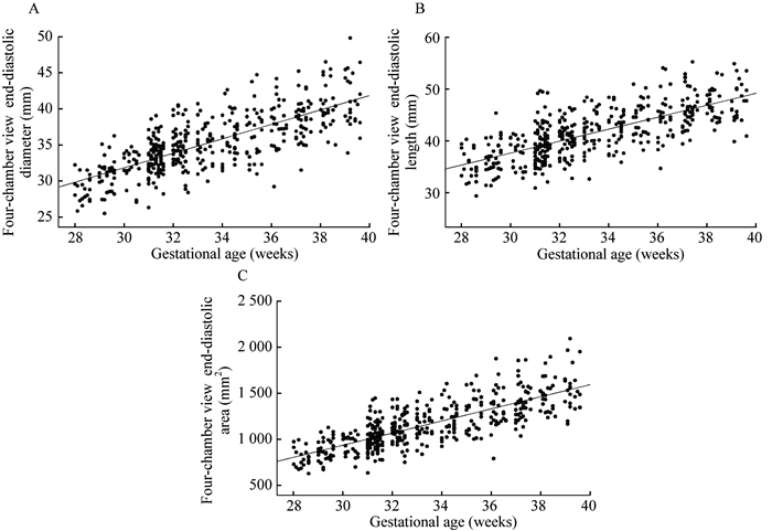
Fig 2
Scatterplots of four-chamber view size with gestational weeks in low-risk fetuses
Tab 1
Reference ranges for end-diastolic diameter, length and area of the four-chamber view
Tab 2
Reference ranges for end-diastolic diameter, length and area of the left ventricle
Tab 3
Reference ranges for end-diastolic diameter, length and area of the right ventricle
Tab 4
Reference ranges for 24-segment spherical index of the left and right ventricles
Tab 5
Left and right ventricular size in low-risk fetuses
($\bar x \pm s$)
Tab 6
Left and right ventricular 24-segmental sphericity index in low-risk fetuses
($\bar x \pm s$)
Tab 7
Interobserver and intraobserver reproducibility for fetal cardiac measurements using fetalHQ